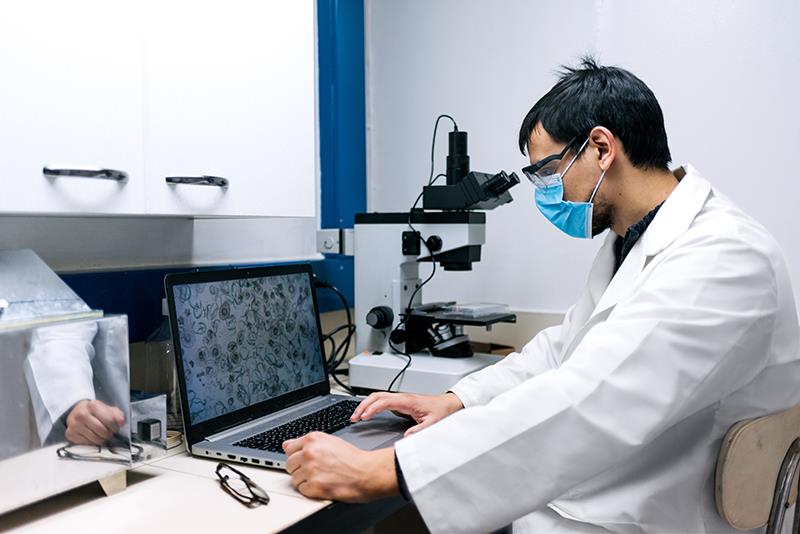
Digital microscopy combined with artificial intelligence (AI) demonstrates comparable accuracy to the reference standard for identifying multiple targets in primary healthcare laboratories and may even increase sensitivity in diagnosis, reports a study.
“With further research addressing challenges such as scalability and cost-effectiveness, AI-supported digital microscopy could improve access to diagnostics, especially in expert-scarce and resource-limited settings,” the researchers said.
A systematic search was performed using the databases of PubMed, Embase, Web of Science, and IEEE. Researchers identified studies that used digital microscopy, AI, and compared results with a standard diagnostic system, with diagnoses performed in a primary healthcare laboratory. Included studies were also published in English, performed on humans, and achieved a sample-level diagnosis.
Two independent researchers conducted the study selection and data extraction, while a third researcher resolved cases of disagreement. The procedure is consistent with the Joanna Briggs Institute methodology for scoping reviews.
Of the 3,403 papers screened, only 22 (0.6 percent) met the eligibility criteria. The following samples were then analysed: blood (n=12) for blood cell and malaria detection, urine (n=4) for urinalysis and parasite detection, cytology of atypical oral (n=1) and cervical cells (n=2), stool (n=2) for parasite detection, and sputum (n=1) for ferning patterns indicating inflammation.
In the sample preparation, both conventional (n=15) and specifically developed methods (n=7) were applied. [J Med Internet Res 2026;28:e78500]
Compared with the reference standard, AI-supported digital microscopy displayed similar diagnostic accuracy for the following tests: complete blood counts, malaria detection, identification of stool and genitourinary parasites, screening for oral and cervical cellular atypia, detection of pulmonary inflammation, and urinalysis.
Notably, sensitivity was greater with AI-support digital microscopy than with manual microscopy in six (85.7 percent) out of seven studies using a reference standard that allowed for this comparison.
“For multiple diagnostic purposes, AI-supported digital microscopy achieved comparable results to the reference standard and could be particularly advantageous for increasing sensitivity in diagnosis,” the researchers said.
Human verification
One study included in this scoping review examined AI-supported digital microscopy with and without human verification. In targets initially classified as positive by the AI models, human verification resulted in a 0.9-percent decrease in sensitivity but a 29.5-percent increase in specificity. [Malar J 2024;23:200]
“This demonstrates that, with human intelligence, AI errors can be identified and removed without a substantial loss of sensitivity,” the researchers said.
“This is in line with the high specificity presented in studies using human verification, which all showed specificity of >90 percent. Expanding human verification to include borderline cases classified as negative may also be used to reduce false negatives and increase sensitivity,” they added. [PLOS Glob Public Health 2024;4:e0003091; PLOS One 2014;9:e104855]
Implications
Potential advantages can be obtained from AI-supported digital microscopy in primary healthcare laboratories relative to manual microscopy, according to the researchers.
For instance, it could enhance diagnostic accuracy, particularly sensitivity, and improve access and timeliness of diagnostics by allowing procedures to be performed at the point-of-care and abolish the need to send samples elsewhere for analysis. [JAMA Netw Open 2021;4:e211740; PLoS ONE 2019;14:e0224885; PLoS Negl Trop Dis 2024;18:e0011967]
Finally, AI-supported digital microscopy could ease the burden of personnel through task shifting. “This could increase the productivity of experts and thereby access to image-based diagnosis,” the researchers said. [Malar J 2024;23:200; PLOS Glob Public Health 2024;4:e0003091]